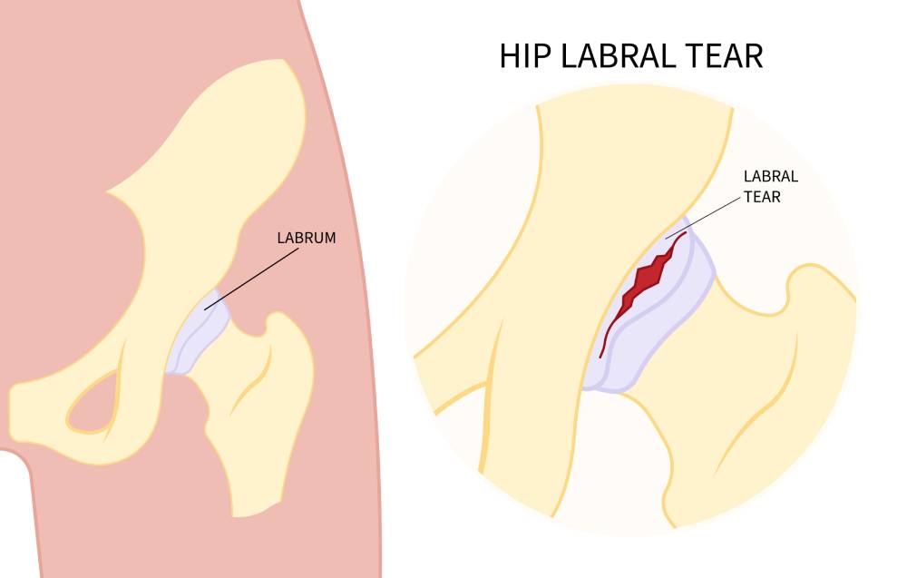
The labrum of the hip joint, which is a ring of cartilage that lines the socket, acts as a cushion and stabilizing structure and keeps the bones tightly in place. Some people are more likely to develop a hip labral tear than others, as it is an injury that occurs gradually over time due to a variation in hip anatomy or repeated trauma.
Anatomy
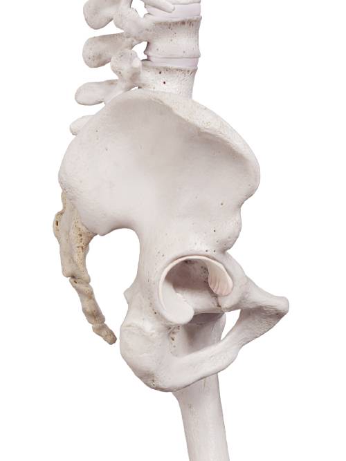
The largest weight-bearing joint in the human body, the hip joint is made of several bones, muscles, and ligaments, and is of the ball and socket variety.
The bones that make up the hip joint are the femur, which is the thigh bone, and the acetabulum on the pelvic bone to which the head of the femur attaches.
The deep concave shape of the acetabulum helps in preventing the femur from dislocating easily. It is surrounded by a joint capsule containing synovial fluid and is stabilized by various muscle tendons and ligaments.
The cartilage of the hip socket it allows the bones to glide smoothly against each other.
The acetabular cartilage extends into softer cartilage called the hip labrum, which increases the depth of the articular socket. By doing this, the labrum allows the hip socket to be more stable and flexible allowing the hip to move through a greater range of movement.
How Does a Hip Labral Tear Occur?
Injury to the hip labrum that covers the acetabulum is commonly referred to as a hip labral tear, which causes problems with day-to-day activities. A lot of cases are caused by trauma, while some may happen over a prolonged time period. It can occur due to:
Trauma
The most common cause of a hip labral tear is trauma to the hip joint. Playing certain sports, such as football, ice hockey, and basketball, can result in labral injury, as repetitive twisting, tackling, pivoting, and other high-collision movements can damage the labrum. Traumatic injury to the pelvic region, such as during a car accident, can also cause a hip labral tear.
Femoroacetabular Impingement (FAI)
Femoroacetabular impingement (FAI) occurs when the femoral head may be not rounded at the top (known as cam impingement), or if the acetabulum has prominent, protruding margins, causing friction between the articular surfaces (pincer deformity), or both.
Femoroacetabular impingement limits flexion at the hip joint and can result in a hip labral tear.
Hip Dysplasia
Hip dysplasia is another condition causing ill-fitted femoroacetabular alignment, in which the socket is too shallow for the femoral head. It occurs due to abnormalities of shape, size and/or orientation of either the head of femur or the socket, or even both. The joint stability is often affected, causing limping, and susceptibility to dislocations. This can also cause a hip labral tear.
Degenerative Conditions
Degeneration of the articular cartilage leads to chronic painful conditions like osteoarthritis. The increased movement of the head of femur and localized pressure within the socket further erodes the cartilage and makes the labrum more susceptible to injury or degeneration. Increasing age can contribute to this and increase the risk of developing osteoarthritis, and subsequently, labral tear.
What Does a Hip Labral Tear Feel Like?
Injury to the hip labrum can cause several symptoms, including:
- Painful and restricted movements of the thigh
- Pain in the groin or the buttocks
- Sensation of clicking or locking felt in the hip during movement
- Worsening of pain after activities such as walking, running, or sitting for prolonged time
- Feeling of stiffness in the hip
- Difficulty and pain during rotating motion
However, it is important to note that in a few cases, hip labral tear might not cause any symptoms at all, at least initially.
Tests for Hip Labral Tear
In case of an injury to the hip labrum, several specialized tests can be performed to aid in the diagnosis. They are often used with imaging tests to come to a diagnosis. These include:
- FABER test – the flexion-abduction-external rotation test can help detect hip joint abnormalities including labral tears.
- McCarthy test – a labral tear is present if passively extending the hip produces a clicking sound
- Fitzgerald test – a collective name given to anterior and posterior labral tear tests; this involves a series of passive motions starting from hip flexion and going on to extension, abduction, external and internal rotation. If the result is pain or a clicking sensation, a hip labral tear is often present
- FADDIR test– passive flexion at hip to 90 degrees, adduction, and then medial rotation; if this movement produces pain, then labral tear is possible
Hip Labral Tear – diagnosis
Injury to the hip labrum can mimic other conditions or can be complicated by pre-existing disorders. To reach a diagnosis, the physician takes a detailed medical history, and performs a physical exam. After this, some tests are performed which provide conclusive evidence, including:
- X-rays – provide a view of the bones, detecting any arthritic changes or bony abnormalities, which might be causing the hip labral tear
- MRI – Magnetic Resonance Imaging gives a visual of the soft tissue surrounding the hip joint, including the labrum. To gain more accuracy, The hip Joint is often injected with a dye to gain a better image of the labrum, this is called an MRI Arthrogram
- MRI 3 Tesla: A higher definition MRI (which reduces the need for an arthrogram and associated discomfort and complications) Research has shown a 3 Tesla MRI to be as accurate as MRI Arthograms in detecting Hip Labral tears
- Diagnostic injection – a painkiller or anesthetic agent is given as an ultrasound-guided injection, and if the pain subsides, a hip labral tear is likely
Treatment
Your options for treatment depend on the cause and severity of injury to the hip labrum, and can include:
Medication
For the management of mild to moderate pain and inflammation in a hip labral tear, some over-the-counter analgesics may be advised, like ibuprofen or other NSAIDs.
Physical Therapy
Certain exercises and muscle stretches promote strengthening and stabilization of the hip joint and improve the range of motion that has become restricted due to labral tear. It is vital to begin these exercises as soon as possible when a labral tear is found.
PRP
Platelet-rich plasma therapy or PRP has proven beneficial in the treatment of several musculoskeletal conditions. Platelets not only help in blood clotting, but PRP also contains several growth factors and other proteins that promote healing and repair of the various tissues in the body. In PRP, the blood is drawn from the patient, and spun through a centrifuge, which separates the platelets. Guided by ultrasound, with the help of a needle, the PRP injection is then injected directly into the hip labral tear.
The process usually takes around 45 to 90 minutes and may cause discomfort due to the repetitive injecting of PRP.
Depending on the degree of injury, PRP sessions are usually performed in a course 3 sessions and is a good alternative to surgery
Prolotherapy
A technique that involves injecting a proliferative substance (that promotes growth of new tissue) into damaged joints to induce healing and repair. Prolotherapy has proven promising in the treatment of hip labral tears. Since cartilage does not have the ability to repair itself, this therapy is helpful for the regeneration of damaged labrum, and can also help avoid surgery.
Surgery
When all the treatment options available have failed to produce effective results, or if the injury is quite severe, surgery is advised. Arthroscopic hip surgery, in which the surgeon works through small incisions, has become increasingly common compared to open hip surgery
Conclusion
Injury to the hip labrum can cause a number of issues and lower the quality of life. Performing daily activities can become difficult and quite painful, and if unresolved can lead to chronic problems and Arthritis. A Hip labral tear can be serious enough to prevent a person from participating in sports, or even walk properly.
Consulting a doctor when the symptoms first start to appear can result in timely intervention, and following a treatment plan that is suitable to your case will help prevent long-term disability and pain.